Contents
Scroll to:
https://doi.org/10.23947/2687-1653-2024-24-3-227-237
EDN: AICZHY
Scroll to:
Introduction. When studying composite materials for construction purposes, it is needed to consider the mechanisms of formation of the structure and properties of modern concretes in the process of strength development. In studies of modern composite materials based on cement binder, there is no information about the development of structural defects and destruction of the material at the initial stages of strength development. This information can be obtained using X-ray computed tomography, a promising method of nondestructive testing of the state of the material. Therefore, the objective of this work was to study the formation and propagation of cracks in samples of fine-grained concrete with different fractional composition of sand due to natural processes of cement shrinkage, as well as the mechanics of destruction of samples of modified fine-grained concrete when applying a compressive load at the early stages of strength development. Materials and Methods. The study used fine-grained concrete mixtures of three compositions with different sand gradation. The tomography samples were made by placing fresh mixtures in polymer cylindrical containers. Tomography of the samples immediately after manufacture, as well as after 8 and 51 days, was performed in a YXLON Cheetah microfocus X-ray machine. The composition with two-fraction sand was modified by mechanical activation of the components, 20×20×20 mm cube samples were made. Further, compression tests were performed at the Instron installation after 3 and 7.5 hours, and then — tomography of the destroyed samples.
Results. It was established that the destruction of contact zones depended on the ratio of the size of the fractions. In the presence of a bulk of coarse sand grains in concrete, the destruction of contact zones was more pronounced and had a main mode. When using fine or polyfraction sand, contact zones were destroyed locally and had a visually smaller area. The images of the destroyed modified sample, tested 3 hours after manufacturing, showed clear cracks and indents on the edges, which indicated the elastic-plastic nature of the destruction. In 7.5 hours, the edges of the sample upon destruction were covered with a network of small cracks; inside the sample there were also numerous cracks and microcracks, which indicated brittle fracture. Based on the obtained images of the deformed structure of modified concrete, the mechanism of transition from elastic-plastic destruction of the material to brittle one was clearly visible.
Discussion and Conclusion. The studied dependences of the influence of the size of fine aggregate on the mechanisms of formation and propagation of structural defects contribute to the theory of the processes of destruction of fine-grained concretes. The results obtained prove the prospects of using X-ray computed tomography as a method of nondestructive testing of the internal structure of fine-grained concrete, including at the early stages of strength development.
Puzatova A.V., Dmitrieva M.A., Tovpinets A.О., Leitsin V.N. Study of Structural Defects Evolution in Fine-Grained Concrete Using Computed Tomography Methods. Advanced Engineering Research (Rostov-on-Don). 2024;24(3):227-237. https://doi.org/10.23947/2687-1653-2024-24-3-227-237. EDN: AICZHY
Introduction. X-ray computed tomography is a promising technique of nondestructive testing of the state of the material. In the concrete industry, tomography is suitable for determining the structure of concrete samples, microcracks, internal fractures, and studying the distribution of pores and aggregate particles. X-ray computed tomography provides the construction of a model of the microstructure of cement paste, allows us to study the development of cement hydration processes [1][2], to make forecasts of the formation of mechanical characteristics and fracture conditions [3][4]. Computed tomography is actively used for studying the average density and porosity of high-strength lightweight concretes [5], the morphology of structural heat-insulating concrete [6], building a mesoscale 3D model of foamed concrete [7], investigating the formation and distribution of pores in lightweight concrete [8], analyzing the microstructural characteristics of concrete samples with various aggregates [9][10], developing three-dimensional mesoscale models for constructing a finite element grid in modeling the structure of concrete [11][12]. In comparison to standard 2D X-ray methods, the construction of 3D models of multicomponent concrete samples is promising for the study of the fundamental mechanisms of formation of the structure and properties of modern concretes.
The most vulnerable area of fine-grained concrete under loading is the contact zone — the contact areas of cement stone and aggregates. When exposed to external loads, it is from these areas that the formation of microdefects and microcracks starts, whose development causes the defect formation at the macro level, which can lead to loss of bearing capacity and structural failure. The destruction of the contact zones occurs due to the difference in characteristics of the bordering components (Young's modulus, Poisson's ratio, thermal linear expansion coefficient, sizes of the contacting phases, microdefects on the interface of the phases) [13]. The X-ray computed tomography method is promising for studying the evolution of contact zones, including at the early stages of hydration. It provides studying the structure without destroying the sample directly during the hardening process. Contact zones, as a rule, have higher porosity and low strength, as a result of which cracks are formed in these zones [4]. The strength of the contact zones is also affected by the size of the aggregate. It has been established that homogeneous and more durable contact zones are formed in concretes with combined aggregates (coarse fraction and crushed) [14]. There are numerous modern studies on the formation of contact zones of cement stone with reinforcement [15], cracks in popcorn concrete [16], defects at the contact boundary of sand-cement mortar with a coarse aggregate [17]. However, along with this, the formation of contact zones in fine-grained concretes with different fineness and packing density of sand grains is poorly studied. In the modern scientific literature, there is no description of the effect of the size of a fine aggregate on the formation of contact zone defects in fine-grained concretes. Thus, the study of the formation of defects in the structure of fine-grained concrete containing sand of aggregate fractional make-up, to obtain a visual picture of crack propagation by X-ray computed tomography is urgent.
To reduce the stresses that occur in the contact zones, microfillers, similar in their properties to cement stone, are used. These fillers, having an increased specific surface area, create additional contact zones between which the stresses arising from the hardening of the binder are redistributed. Hardening of the contact zones can be achieved through introducing mechanically activated components into the concrete mixture [13]. Mechanical activation of separate components contributes to the formation of a denser structure, the preservation of uniformity in composition, the development of initial strength due to the acceleration of the hydration reaction and the growth of crystallohydrates of cement stone, as well as the reduction in the setting time [18][19]. The development of structural defects in fine-grained concretes modified by mechanical activation of components has also been poorly studied. Among modern scientific papers, there are very few works devoted to the study of crack propagation processes in the modified structure of fine-grained concretes at the initial stages of strength development. Therefore, the use of the computed tomography method to study the mechanics of destruction of samples of modified fine-grained concrete at the initial stages of hardening is important today.
This work was aimed at studying the formation and propagation of cracks in samples of fine-grained concrete with different fractional make-up of sand due to natural processes of shrinkage of cement stone, as well as the mechanics of destruction of samples of modified fine-grained concrete when applying a compressive load at the early stages of strength development.
Materials and Methods. In the research, to determine the defects of the contact zones of fine-grained concretes with different sand gradation, three samples of fine-grained concrete mixture of the following compositions were used:
Freshly-mixed compositions of fine-grained concrete mix were placed in a polymer cylindrical container with a diameter of 8 mm and a length of about 70 mm. The diameter of the container was determined in accordance with the size of the initial components of fine-grained concrete of the previously mentioned compositions based on the conditions of representativeness [20], and representativeness of the volume under study. To track changes in the structure under cement hydration and cement stone shrinkage, tomography of the samples was performed immediately after mixing the components and after 8 days of hardening. Samples at the age of 51 days were taken as the final result.
In the study on the evolution of the crack formation mechanism during the destruction of samples under the action of an external compressive force, composition No. 3 with two-fraction sand, modified by mechanical activation of the cement and sand composition, was used. Mechanical activation of the components (cement and sand) was carried out using a high energy ball mill Retsch EMax. The components were crushed at a rotation speed of the apparatus bowls of 750 rpm for 5 minutes. Cubic samples of 20×20×20 mm in size were made from concrete mortars, to which an external compressive load was applied after 3 and 7.5 hours. After applying the load, tomography of the destroyed samples was performed to trace the evolution of the fracture mode of the material.
The structure of the concrete samples was studied using a YXLON Cheetah microfocus X-ray computed tomograph with Y. Cheetah configuration. The device specifications are shown in Table 1.
Table 1
YXLON Cheetah Tomograph Specifications
|
Characteristic |
Value |
|
X-ray tube |
Open-type |
|
Operating voltage range, kV |
25–160 |
|
Operating current range, mA |
0.1–1.0 |
|
Maximum tube power, W |
64 |
|
Maximum tube power on target, W |
15 |
|
Detector tilt angle |
±70о (segment 140°) |
|
Magnification (geometric/maximum) |
2000х/17500х |
|
Maximum sample dimensions, mm |
800х500 |
|
Time from sample loading to first image, sec |
<10 |
|
Time for full tomography of the sample, sec |
7 |
|
Time for laminography of the sample, sec |
20 |
|
Overall dimensions, mm |
1650х1400х1850 |
|
Weight, kg |
2,200 |
From the perspective of micromechanics of composite materials, the assessment of effective characteristics can be introduced by sets of properties of a representative volume of the body under study. The test sample for tomography should correspond to a representative volume of material, which makes sense of the elementary macrovolume of a microinhomogeneous medium.
During the experiment, after X-rays pass through the sample, a set of flat X-ray images with an uneven distribution of grayness in the images is obtained. This is due to the uneven absorption of X-rays by the components of the studied material — the presence of pores, defects, dense inclusions, etc. After reconstruction of 3D images of the sample, the grayness gradient is inverted relative to conventional X-ray images: the materials that are most transparent to X-rays, e.g., pores, correspond to black, and the densest material is white. The grayness density in full-color representation is considered in the range 0–255, where 0 corresponds to black, and 255 corresponds to white. This algorithm is used to determine the minimum size of features that could be differentiated as structural components of the sample under study: porosity, cement mortar, and aggregate in volume. Further, using this grayscale, highlighting certain numerical ranges, it is possible to analyze a separate internal structure, distribution of components and porosity [21].
The shooting parameters for all the studied samples remained constant: voltage — 85 kW; current — 45 µA; approximation — 8.9; scanning angle — 360°. The survey results represent 1,024 consecutive images of the internal structure of the samples. Further processing of the resulting array of two-dimensional images took place in the “Volume Graphics Studio” program. To improve the visualization of inclusions, editing layered images by brightness and contrast levels was performed. The result of the tomography was a 3D model of the sample and its three projections with the possibility of studying the internal structure in any section.
Research Results. Images of the internal structure of samples of composition No. 1 with monofraction sand (fraction 0.63–0.315 mm) at the age of 0, 8, and 51 days obtained by X-ray tomography are shown in Figure 1. The darkest areas in the images have the lowest density, in this case, the pores. The hardest particles correspond to the lightest areas.
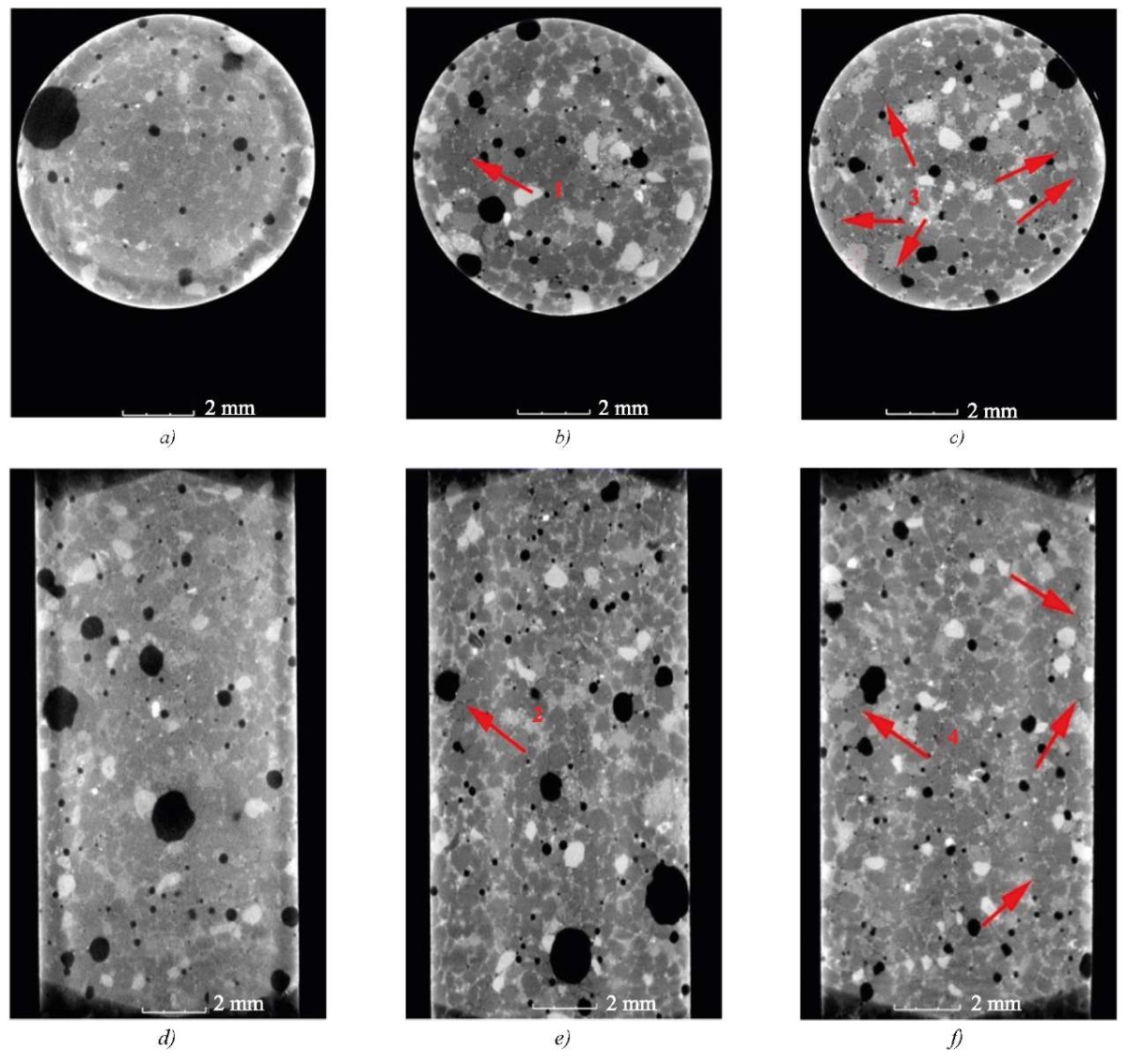
Fig. 1. Internal structure of sample No. 1 (monofraction sand):
a, d — immediately after preparation;
b, f — at the age of 8 days;
c, f — at the age of 51 days
The images of the internal structure of the sample of composition No. 1 with monofraction sand showed no changes in the contact zones immediately after mixing. By the 8th and 51st days of hardening, dark stripes were visible around separate sand grains (indicated by red arrows), corresponding to voids that were formed due to shrinkage of the cement stone. Moreover, with the increasing age of concrete, such voids around the sand grains visually became more numerous. Enlarged images of separate fractured voids are shown in Figure 2.
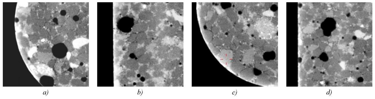
Fig. 2. Enlarged fragments of Figure 1:
a — fragment 1; b — fragment 2; c — fragment 3; d — fragment 4
In fragments 3 and 4 corresponding to sample No. 1 at the age of 51 days, the mode of the manifestation of defects in the contact zones around sand particles was most pronounced. This was confirmed by the fact that by 51 days, the process of shrinkage of cement stone was almost complete, whereas at the age of 8 days, the shrinkage was in the active phase.
Images of the internal structure of samples of composition No. 2 with polyfraction sand at the age of 0, 8 and 51 days are shown in Figure 3. Separate enlarged fragments are shown in Figure 4.
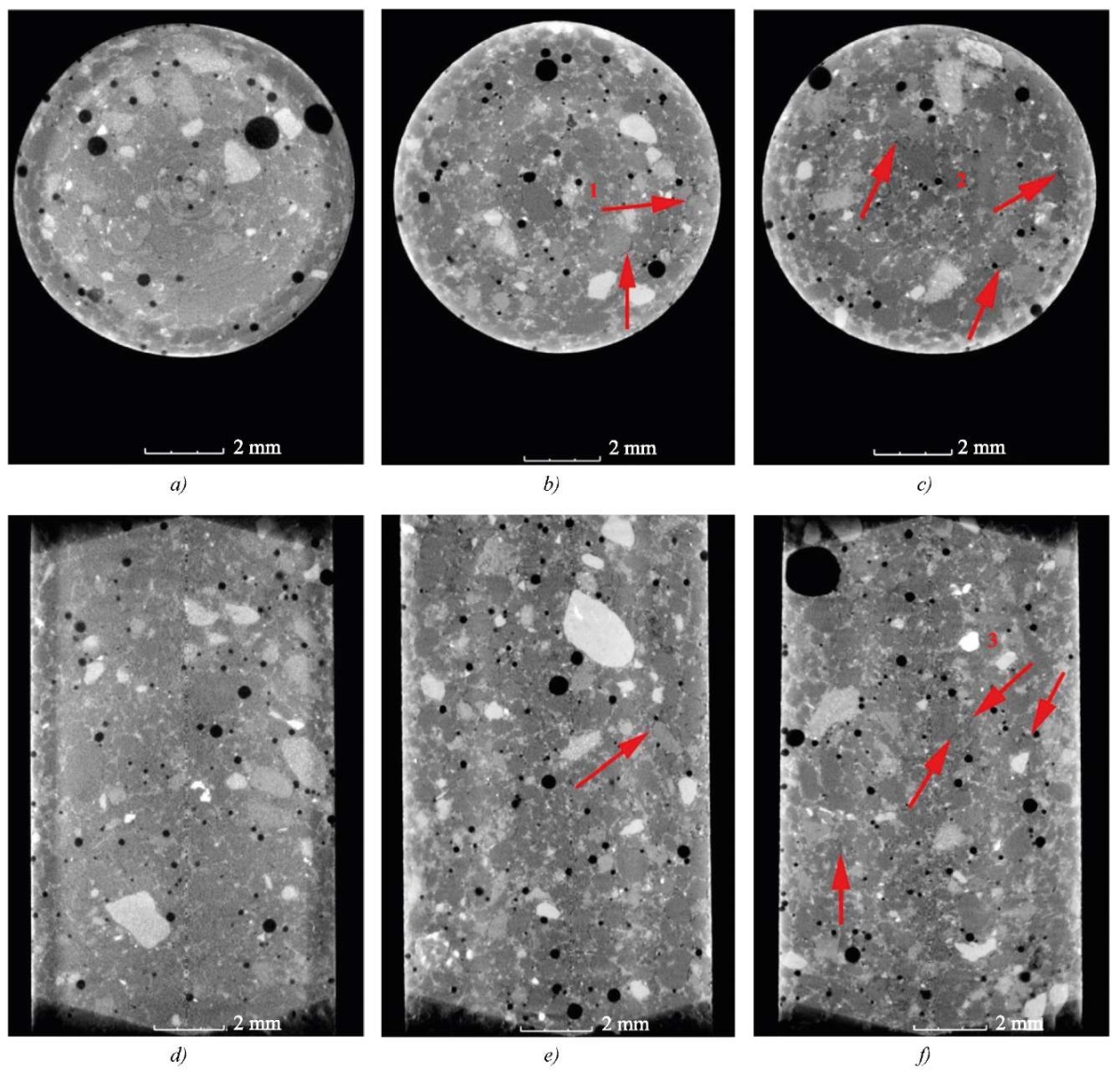
Fig. 3. Internal structure of sample No. 2 (polyfraction sand):
a, d — immediately after preparation;
b, e — at the age of 8 days;
c, f — at the age of 51 days
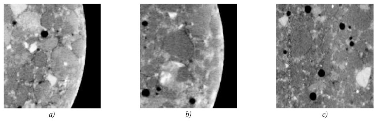
Fig. 4. Enlarged fragments of Figure 3:
a — fragment 1; b — fragment 2; c — fragment 3
The development of defects in the contact zones of samples of composition No. 2 with a polyfraction aggregate, as in composition No. 1 with a monofraction aggregate, manifested itself by the 8th day of hardening, the number of defective areas increased by the 51st day. It can be noted that the destruction of contact zones around individual large sand particles is not observed.
Images of the internal structure of samples of composition No. 3 with two-fraction sand (fractions 2.5–1.25 mm and 0.63–0.315 mm, no intermediate fraction) at the age of 0, 8, and 51 days are shown in Figure 5.
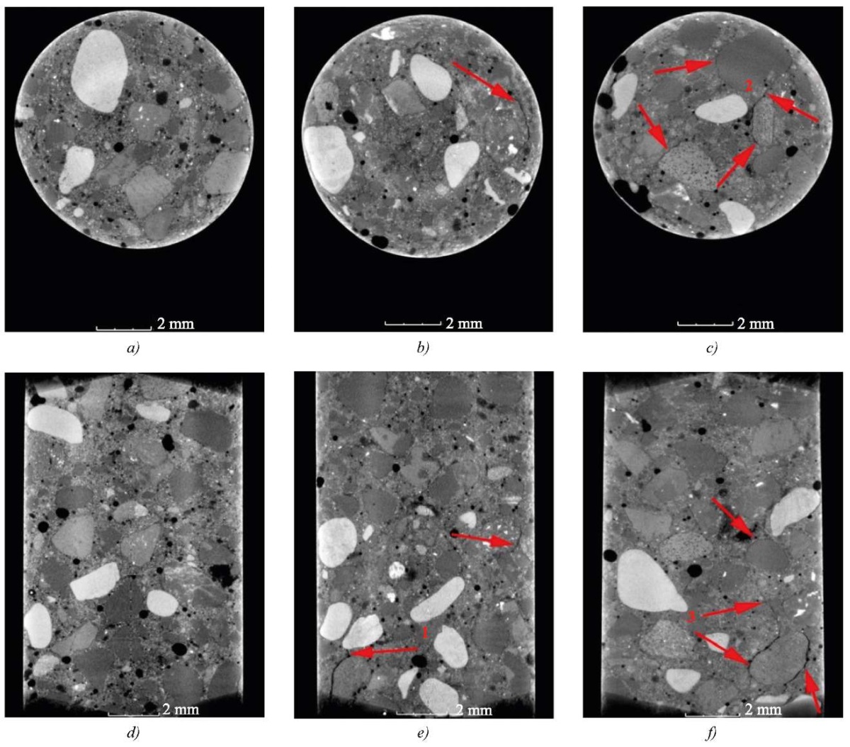
Fig. 5. Internal structure of sample No. 3 (two-fraction sand):
a, d — immediately after preparation;
b, e — at the age of 8 days;
c, f — at the age of 51 days
In the images of the internal structure of the samples containing two fractions of sand, at the age of 8 and 51 days, a clear formation of cracks around large grains of sand was observed, and the main mode of the crack formation was traced when the cracks were connected to each other (Fig. 6).
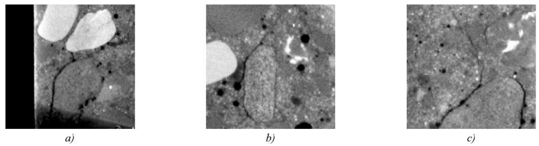
Fig. 6. Enlarged fragments of Figure 5:
a — fragment 1; b — fragment 2; c — fragment 3
The formation of main cracks near coarse sand grains indicated that the contact zones around coarse aggregate particles were most stressed and susceptible to destruction during the shrinkage of the cement stone.
Based on the obtained images of the internal structure of samples with different sand sizes, it can be concluded that the development of contact zone defects due to shrinkage of the cement stone depends on the ratio of fraction sizes. In samples with mono- and polyfraction sand, the defective structure develops locally, the area of such defects is visually much smaller than in samples with two fractions of different sizes. In the presence of a coarse fraction of sand with a high-volume concentration, defects in contact zones develop near coarse grains and have the main mode.
Cube samples of composition No. 3 with two-fraction sand, modified by mechanical activation of individual components, were subjected to the application of external compressive load at the age of 3 and 7.5 hours after preparation. Images of the deformed internal structure of the samples are shown in Figure 7.
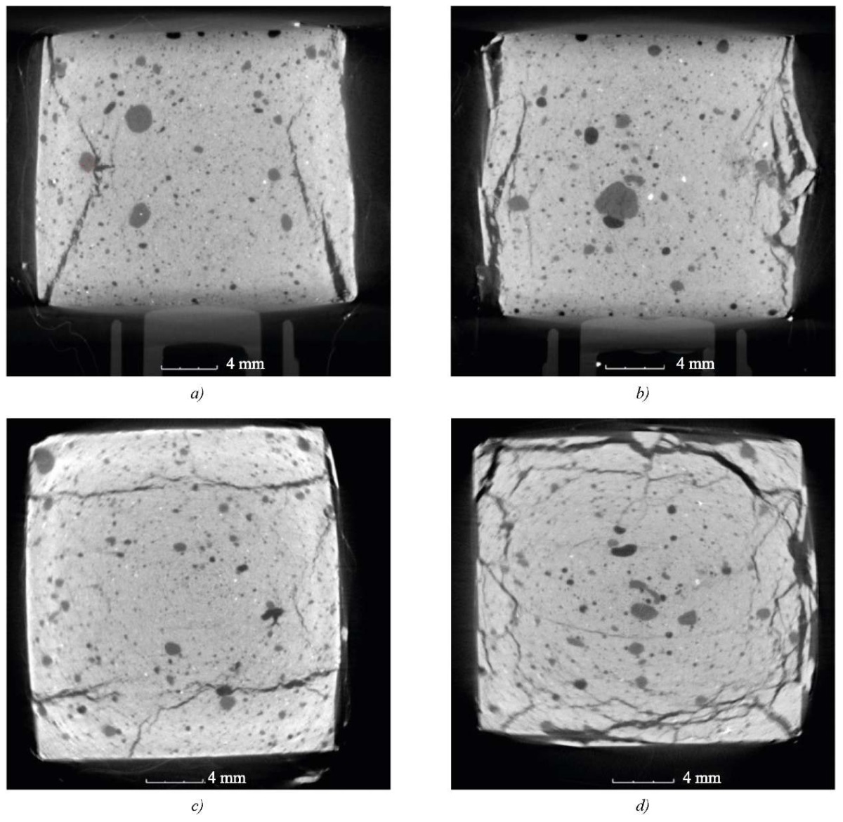
Fig. 7. Images of the internal structure of modified samples:
a — frontal section at the age of 3 hours;
b — frontal section at the age of 7.5 hours;
c — horizontal section at the age of 3 hours;
d — horizontal section at the age of 7.5 hours
The images showed the evolution of hardening concrete samples from elastic-plastic to brittle fracture. The samples tested 3 hours after production had an elastic-plastic fracture pattern, with clear cracks and chips on the sample edges. At the age of 7.5 hours, the sample edges were covered with a network of small cracks during fracture, and there were also numerous cracks and microcracks inside the sample, indicating brittle fracture.
Discussion and Conclusion. Thus, by means of mechanical tests and X-ray computed tomography, it is possible to track the processes of destruction in the structure of fine-grained concrete. The results obtained give rise to a new complex method for assessing the structural characteristics of modified fine-grained concrete at all stages of strength development.
It has been established that the destruction of contact zones depends on the ratio of fraction sizes. In the bulk of coarse sand particles in the body of concrete, the destruction of contact zones is more pronounced and has the main mode. When using fine or polyfraction sand, contact zones are destroyed locally and have a visually smaller area. This indicates that contact zones near coarse aggregate particles are most stressed and are primarily subject to destruction under the shrinkage of cement stone.
The studied dependences of the influence of the fine aggregate sizes on the mechanisms of formation and propagation of structural defects contribute to the theory of destruction processes of fine-grained concrete. The results obtained prove the prospects of using X-ray computed tomography as a method of non-destructive testing of the internal structure of fine-grained concrete, including at the early stages of strength development. Computer tomography, along with traditional methods of investigation of the structure and properties of building materials, gives rise to a new complex method that provides studying modern multicomponent concretes at all stages of strength development, the mechanisms of formation and propagation of structural defects due to natural processes of changing the state of the material and under various external loading conditions.
1. Dmitrieva MA, Kogai AD, Leitsin VN, Tovpinets AO, Shinyaeva MV. An Experimental and Theoretical Approach to Assessing the Structure of Fine-Grained Modified Concretes. Vestnik MGSU. 2023;18(1):70–81. https://doi.org/10.22227/1997-0935.2023.1.70-81
2. Dmitrieva MA, Sharanova AV, Leitsin VN, Shinyaeva MV. Experimental Studies of the Evolution of Structural and Mechanical Characteristics of Cement Stone in the Process of Hydration. In: Proc. III International Conference “Advanced Building Materials and Technologies”. Kaliningrad: IKBFU Publ.; 2021. P. 7–13. (In Russ.)
3. Mingzhong Zhang, Jivkov AP. Micromechanical Modelling of Deformation and Fracture of Hydrating Cement Paste Using X-ray Computed Tomography Characterization. Composites Part B: Engineering. 2016;88:64–72. https://doi.org/10.1016/j.compositesb.2015.11.007
4. Lavrov A, Panduro EAC, Torsæter M. Synchrotron Study of Cement Hydration: Towards Computed Tomography Analysis of Interfacial Transition Zone. Energy Procedia. 2017;114:5109–5117. https://doi.org/10.1016/j.egypro.2017.03.1666
5. Inozemtsev AS. Average Density and Porosity of High-Strength Lightweight Concrete. Magazine of Civil Engineering. 2014;51(7):31–37. (In Russ.) https://doi.org/10.5862/MCE.51.4
6. Osipov SP, Prishchepa IA, Kudyakov AI, Batranin AV, Osipov OS. Computer Tomography of Foam Concrete. Systems. Methods. Technologies. 2018;38(2):146–152. https://doi.org/10.18324/2077-5415-2018-2-146-152
7. Tuan Nguyen, Abdallah Ghazlan, Alireza Kashani, Stćphane Bordas, Tuan Ngo. 3D Meso-Scale Modelling of Foamed Concrete Based on X-ray Computed Tomography. Construction and Building Materials. 2018;188:583–598. https://doi.org/10.1016/j.conbuildmat.2018.08.085
8. Haizhu Lu, Eugene Alymov, Sanjay Shah, Karl Peterson. Measurement of Air Void System in Lightweight Concrete by X-ray Computed Tomography. Construction and Building Materials. 2017;152:467–483. https://doi.org/10.1016/j.conbuildmat.2017.06.180
9. Khuzin AF, Rahimov RZ. The Effect of Multiwalled Carbon Nanotubes on the Porosity of the Cement Stone. News KSUAE. 2016;37(3):231–237.
10. Sang-Yeop Chung, Mohamed Abd Elrahman, Dietmar Stephan, Paul H Kamm. The Influence of Different Concrete Additions on the Properties of Lightweight Concrete Evaluated Using Experimental and Numerical Approaches. Construction and Building Materials. 2018;189:314–322. https://doi.org/10.1016/j.conbuildmat.2018.08.189
11. Yujie Huang, Zhenjun Yang, Wenyuan Ren, Guohua Liu, Chuanzeng Zhang. 3D Meso-Scale Fracture Modelling and Validation of Concrete Based on In-situ X-ray Computed Tomography Images Using Damage Plasticity Model. International Journal of Solids and Structures. 2015;67–68:340–352. https://doi.org/10.1016/j.ijsolstr.2015.05.002
12. Levandovskiy AN, Melnikov BE, Shamkin AA. Porous Material Modeling with Finite Element Method. Construction of Unique Buildings and Structures. 2017;53(2):61–77. https://doi.org/10.18720/CUBS.53.5
13. Bolshakov VI, Yelisieieva MO, Shcherbak SA. Contact Strength of Mechanoactivated Fine Concretes from Granulated Blast-Furnace Slags. Nauka ta progres transportu. 2014;53(5):138–149. (In Russ.)
14. Egorochkina IO, Serebryanaya IA, Shlyakhova EA, Matrosov AA, Pronina KA, Kuzina AN. Study of the Structure of the Contact Zone in Concretes with Combined Aggregates. Engineering Journal of Don. 2019;55(4):40.
15. Bedarev VV, Bedarev NV, Bedarev AV. The Destruction of Concrete in the Contact Layer Based on the Basic Provisions of General Theory of Adhesion and Anchoring of Periodic Profile Reinforcement in Concrete. In: Proc. International Construction Congress “Science. Innovations. Goals. Construction”. Moscow: Research Center of Construction; 2023. P. 39–43. (In Russ.) https://doi.org/10.37538/2949-219%D0%A5-2023-39-43
16. Pichugin AP, Khritankov VF, Smirnova OE, Pimenov EG. Crack Formation in Large-Porous Concrete with an Integral Arrangement of a Large Filler. Expert: Theory and Practice. 2020;7(4):47–52. (In Russ.)
17. Pschenichniy GN. About the Features of Cement Concrete Surface Area. Concrete Technologies. 2015;110–111(9–10):56–60.
18. Dmitrieva MA, Sharanova AV, Panfilova AD, Plakhtiy AA. Rheological Properties of Building Mortars Used for 3D Printing. In: Proc. International Conference “Advanced Building Materials and Technologies”. Kaliningrad: IKBFU Publ.; 2019. P. 18–32. (In Russ.)
19. Sharanova AV, Dmitrieva MA, Leitsin VN. Ensuring Formation of Concrete Building Constructions without Formwork by Additive Technologies. In: Proc. II International Conference “Advanced Building Materials and Technologies”. Kaliningrad: IKBFU Publ.; 2020. P. 15–21.
20. Dvorak G.J. Micromechanics of Composite Materials. Series: Solid Mechanics and Its Applications. Dordrecht: Springer; 2013. 442 p. https://doi.org/10.1007/978-94-007-4101-0
21. Leitsin VN, Dmitrieva MA, Ivonin IV, PonomarevSV, Polyushko VA, Tovpinets AO, et al. Determining Factors ofthe Formation of the Structure of Low-Temperature Ceramics. Physical Mesomechanics. 2017;20(6):77–85. (In Russ.)
Anastasiia V. Puzatova, Head of the Laboratory of Construction Materials, Senior Lecturer of the Education and Research Cluster, Institute of High Technology
14, Alexander Nevsky Str., Kaliningrad, 236041
Maria A. Dmitrieva, Dr.Sci. (Phys.-Math.), Professor of the Education and Research Cluster, Institute of High Technology
14, Alexander Nevsky Str., Kaliningrad, 236041
Alexandr O. Tovpinets, Researcher of the Education and Research Cluster, Institute of High Technology
14, Alexander Nevsky Str., Kaliningrad, 236041
Vladimir N. Leitsin, Dr.Sci. (Phys.-Math.), Professor of the Education and Research Cluster, Institute of High Technology
14, Alexander Nevsky Str., Kaliningrad, 236041
Puzatova A.V., Dmitrieva M.A., Tovpinets A.О., Leitsin V.N. Study of Structural Defects Evolution in Fine-Grained Concrete Using Computed Tomography Methods. Advanced Engineering Research (Rostov-on-Don). 2024;24(3):227-237. https://doi.org/10.23947/2687-1653-2024-24-3-227-237. EDN: AICZHY

Advanced Engineering Research (Rostov-on-Don)
ISSN 2687-1653 (Online)
Contact with: Publisher / Editorial Office of the Journal
Publisher: Don State Technical University - DSTU, Rostov-on-Don, Russia - https://donstu.ru/en/
Editor-in-Chief: Alexey N. Beskopylny, Dr.Sci. (Eng.), Professor, Vice-Rector, Don State Technical University (Rostov-on-Don, Russia)
Don State Technical University
1, Gagarin Sq., Rostov-on-Don, 344003, Russia
tel.: +7 (863) 2738-372, e-mail: vestnik@donstu.ru
16+
Processing of personal data